Lyme is the most common vector-borne disease in the United States, according to the CDC, and it’s primarily caused by a bacteria called Borrelia burgdorferi. B. burgdorferi is transmitted from a tick bite and accompanied by symptoms, such as headaches, fatigue, aches and joint pain. When examining why joint pain occurs in Lyme disease, there are many possible sources such as molecular mimicry, a process where the immune system creates antibodies to certain tissue amino acid sequences which are similar to self-tissues. Inflammation in general can also cause joint pain. Moreover, an interesting and significant source of join pain includes the complex impacts Lyme has on connective tissue.
Lyme’s Impacts on Connective Tissue
Borrelia can break down soluble and insoluble ground substances within the extracellular matrix1 and also activate metalloproteases.2 This causes collagen to dissolve, allowing B. burgdorferi to begin colonizing the collagen fibers. Interestingly, Borrelia can also delay the tissue healing process because it inhibits the regeneration of collagen by fibronectin.3 This mechanism is not distinct to Lyme; it has been postulated that Mycoplasma is a potential culprit in rheumatoid arthritis (RA) as well. One study in the Journal of Clinical Microbiology detected Mycoplasma fermentans in 88% of cases of RA.4
How to Address Collagen Damage and Loss in Lyme
In functional medicine, addressing a symptom always involves determining how it originated in order to target the root cause. In the case of collagen degradation in Lyme, it is imperative to address B. burgdorferi and any co-infections as the root cause. Additionally, supporting a patient’s collagen-rich areas of the body, such as skin, joints, vessels, and eyes, can be vital while addressing the infection.
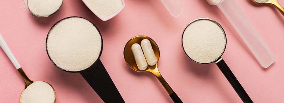
Nutrient-dense dietary choices, stress management, sleep optimization and avoiding toxins all aid in protecting collagen, but patients may also benefit from supplemental collagen. While some types of supplemental collagen are available in pill form, patients can also conveniently add powder formulations to beverages to support their bodies as they keep up with collagen repair.
Four Key Ingredients to Look for in a Collagen Supplement
Collagen Hydrolysate
This ingredient boasts short-chain peptides with a molecular weight ideal for maximum absorption in the body.5-6 In research, collagen hydrolysate has been found to stimulate chondrocytes to produce type II collagen and aggrecan, which are the glue of the extracellular matrix.
Hyaluronic Acid Extract
This ingredient provides ideal support for the viscoelastic, lubricating properties of synovial fluid, which reduces friction in joints. Studies on hyaluronic acid extract suggest it can also significantly increase synovial fluid production and prevent its degradation.8
Type I Collagen and Mucopolysaccharides
Type I collagen and mucopolysaccharides provide support for tendons, ligaments and fascial tissue. Mucopolysaccharides add to the stretch and resilience of tendons and ligaments as well as support proper tendon fiber healing.9
Magnesium and Vitamin C
Magnesium is often depleted in musculoskeletal tissue, leading to soreness, and vitamin C is a vital cofactor for the gradual crosslinking of collagen and connective tissue.
The Bottom Line
When connective tissue repair or regeneration is needed, our bodies rely on type II collagen and extracellular matrix material, like hyaluronic acid and other polysaccharides. These components help aggregate, organize and heal connective tissue fiber to maximize strength, mobility and elasticity. Without these components, healing can instead result in type III collagen, which is randomly oriented, immature and weak. Proper nutritional support for your patients can reduce rehabilitation time and stimulate the collagen they need to ease joint pain.
Angela Lucterhand, DC

Dr. Angela Lucterhand received her doctorate in chiropractic from Palmer College of Chiropractic in Davenport, Iowa. While there, she was a graduate teaching assistant for courses including Anatomy and Physiology, Histology, the Central Nervous System, and Spinal Anatomy. She received her Bachelor of Arts at Indiana University.
Her interest in basic health care led her to become a Health Education Specialist with the Peace Corps, where she served in Mali, West Africa. Dr. Lucterhand loves teaching and was an adjunct professor for Anatomy and Physiology at Ivy Tech. Her passion in practice is addressing the root causes of disease and emphasizing the impact of lifestyle medicine.
References
- Coleman, J. L., Roemer, E. J., & Benach, J. L. (1999). Plasmin-coated borrelia Burgdorferi degrades soluble and insoluble components of the mammalian extracellular matrix. Infection and Immunity, 67(8), 3929–3936. https://doi.org/10.1128/IAI.67.8.3929-3936.1999
- Zambrano, M.C., Beklemisheva, A.A., Bryksin, A.V., Newman, S.A., & Cabello, F.C. (2004). Borrelia burgdorferi binds to, invades, and colonizes native Type I collagen Lattices. Infection and Immunity, 72(6), 3138–3146. https://doi.org/10.1128/IAI.72.6.3138-3146.2004
- Grab, D.J., Lanners, H.N., Martin, L.N., Chesney, J., Cai, C., Adkisson, H.D., & Bucala, R. (1999). Interaction of Borrelia burgdorferi with Peripheral Blood Fibrocytes, Antigen-Presenting Cells with the Potential for Connective Tissue Targeting. Molecular Medicine 5, 46–54. https://doi.org/10.1007/BF03402138
- Johnson, S., Sidebottom, D., Bruckner, F., & Collins, D. (2000). Identification of Mycoplasma fermentans in synovial fluid samples from arthritis patients with inflammatory disease. Journal of Clinical Microbiology, 38(1), 90–93. https://doi.org/10.1128/JCM.38.1.90-93.2000
- Iwai, K., Hasegawa, T., Taguchi, Y., Morimatsu, F., Sato, K., Nakamura, Y., Higashi, A., Kido, Y., Nakabo, Y., & Ohtsuki, K. (2005). Identification of food-derived collagen peptides in human blood after oral ingestion of gelatin hydrolysates. Journal of Agricultural and Food Chemistry, 53 16, 6531-6.
- Oesser, S., Adam, M., Babel, W., & Seifert, J. (1999). Oral administration of (14)C labeled gelatin hydrolysate leads to an accumulation of radioactivity in cartilage of mice (C57/BL). The Journal of Nutrition, 129 10, 1891-5.
- Lundquist, P. & Artursson, P. (2016). Oral absorption of peptides and nanoparticles across the human intestine: Opportunities, limitations and studies in human tissues. Advanced Drug Delivery Reviews, 106. doi: 10.1016/j.addr.2016.07.007
- Möller, I., Martínez-Puig, D., & Chetrit, C. (2009). LB021 Oral Administration of a Natural Extract Rich in Hyaluronic Acid for the Treatment of Knee OA with Synovitis: A Retrospective Cohort Study. Clinical Nutrition Supplements, 4, 171-172.
- Shakibaei, M., Buhrmann, C., & Mobasheri, A. (2011). Anti-inflammatory and anti-catabolic effects of TENDOACTIVE® on human tenocytes in vitro. Histology and Histopathology, 26 9, 1173-85.


